Aphthous Stomatitis
Diagnosis
Most people affected by occasional minor aphthous ulceration do not require tests. They are undertaken if there are recurrent attacks of multiple or severe oral ulcers or complex aphthosis.

Blood tests may include:
- Blood count, iron, B12 and folate studies
- Gluten antibody tests for coeliac disease
- Faecal calprotectin test for Crohn disease
- Swabs for microbiology evaluate the presence of Candida albicans, Herpes simplex virus and Vincent's organisms.
Other causes of mouth ulcer should be considered, including:
- Herpes simplex
- Herpangina
- Hand, foot and mouth disease
- Erythema multiforme
- Fixed drug eruption
Appearance
Diagnosis is mostly based on the clinical appearance and the medical history. The most important diagnostic feature is a history of recurrent, self healing ulcers at fairly regular intervals.
Although there are many causes of oral ulceration, recurrent oral ulceration has relatively few causes, most commonly aphthous stomatitis, but rarely Behçet's disease, erythema multiforme, ulceration associated with gastrointestinal disease, and recurrent intra-oral herpes simplex infection.
Special investigations

Special investigations may be indicated to rule out other causes of oral ulceration. These include blood tests to exclude anemia, deficiencies of iron, folate or vitamin B12 or celiac disease. However, the nutritional deficiencies may be latent and the peripheral blood picture may appear relatively normal.
Some suggest that screening for celiac disease should form part of the routine work up for individuals complaining of recurrent oral ulceration. Many of the systemic diseases cause other symptoms apart from oral ulceration, which is in contrast to aphthous stomatitis where there is isolated oral ulceration.
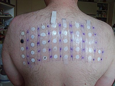
Patch testing may be indicated if allergies are suspected (e.g. a strong relationship between certain foods and episodes of ulceration). Several drugs can cause oral ulceration (e.g. nicorandil), and a trial substitution to an alternative drug may highlight a causal relationship.
Biopsy
Tissue biopsy is not usually required, unless to rule out other suspected conditions such as oral squamous cell carcinoma. The histopathologic appearance is not pathognomonic (the microscopic appearance is not specific to the condition).
Early lesions have a central zone of ulceration covered by a fibrinous membrane. In the connective tissue deep to the ulcer there is increased vascularity and a mixed inflammatory infiltrate composed of lymphocytes, histiocytes and polymorphonuclear leukocytes.

